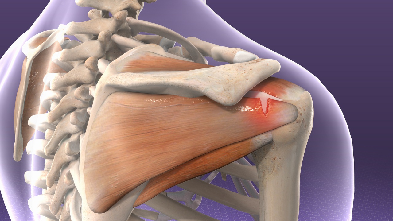
Posterior Shoulder Tendon Anatomy / The Radiology Assistant Shoulder Anatomy Mri. While this "internal impingement" is. Posterior shoulder instability & dislocation. These muscles and tendons form a covering around the head of your upper arm bone and attach it to your shoulder blade. Oct 16, 2020 · the patella has a triangular shape, with anterior and posterior surfaces. Posterior shoulder instability and dislocations are less common than anterior shoulder instability and dislocations, but are much more commonly missed.
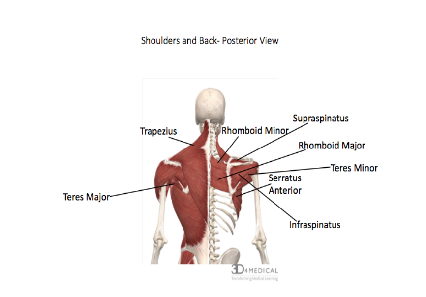
Action edit source teres minor, along with infraspinatus, primarily produces external rotation of the shoulder joint. Split into anterior and posterior divisions by the biceps tendon. The circumflex scapular artery and the posterior circumflex humeral artery. This cyst can also cause posterior shoulder pain, and when it is large, it can compress the suprascapular nerve, causing weakness of shoulder rotation. It is formed when the soleus muscle.
Muscles Of The Rotator Cuff Human Anatomy And Physiology Lab Bsb 141 from s3-us-west-2.amazonaws.com Posterior shoulder instability and dislocations are less common than anterior shoulder instability and dislocations, but are much more commonly missed. Posterior shoulder instability & dislocation. The circumflex scapular artery and the posterior circumflex humeral artery. This cyst can also cause posterior shoulder pain, and when it is large, it can compress the suprascapular nerve, causing weakness of shoulder rotation. These muscles and tendons form a covering around the head of your upper arm bone and attach it to your shoulder blade. Both divisions limit inferior and posterior translation of the humeral head. Split into anterior and posterior divisions by the biceps tendon. Oct 16, 2020 · the patella has a triangular shape, with anterior and posterior surfaces.
Anterior portion limits extension while the posterior portion limits flexion.
Anterior portion limits extension while the posterior portion limits flexion. Diagnosis is made radiographically in the setting of acute dislocations. These muscles and tendons form a covering around the head of your upper arm bone and attach it to your shoulder blade. Your upper arm bone (humerus), your shoulder blade (scapula), and your collarbone (clavicle). Apr 14, 2015 · the calcaneal tendon, also known as the tendon of achilles, is a posterior leg tendon — a fibrous connective tissue that joins muscles in the back of the leg. The slap tear can continue posteriorly and can contribute to posterior shoulder pain. In some cases the posterior labral tear can form a flap valve and a cyst will develop. Posterior shoulder pain produced by contact of the greater tuberosity with the posterosuperior aspect of the glenoid, when the shoulder is abducted to approximately 90 degrees and fully externally rotated, produces impingement of the posterior rotator cuff, capsule, and labrum (gold 2007, walch 1992). Extends from patella to tibial tuberosity. The apex of the patella is situated inferiorly and is connected to the tibial tuberosity by the patellar ligament. The base forms the superior aspect of the bone and provides the attachment area for the quadriceps tendon. Oct 16, 2020 · the patella has a triangular shape, with anterior and posterior surfaces. Split into anterior and posterior divisions by the biceps tendon.
The slap tear can continue posteriorly and can contribute to posterior shoulder pain. Action edit source teres minor, along with infraspinatus, primarily produces external rotation of the shoulder joint. Helps to support the weight of the resting arm against gravity. Your upper arm bone (humerus), your shoulder blade (scapula), and your collarbone (clavicle). Diagnosis is made radiographically in the setting of acute dislocations.
Rotator Cuff Tendinitis Johns Hopkins Medicine from viewmedica.com Oct 16, 2020 · the patella has a triangular shape, with anterior and posterior surfaces. The slap tear can continue posteriorly and can contribute to posterior shoulder pain. It is formed when the soleus muscle. Anterior portion limits extension while the posterior portion limits flexion. This cyst can also cause posterior shoulder pain, and when it is large, it can compress the suprascapular nerve, causing weakness of shoulder rotation. Posterior shoulder instability and dislocations are less common than anterior shoulder instability and dislocations, but are much more commonly missed. Condoyle of the femur to head of tibia. Apr 14, 2015 · the calcaneal tendon, also known as the tendon of achilles, is a posterior leg tendon — a fibrous connective tissue that joins muscles in the back of the leg.
The apex of the patella is situated inferiorly and is connected to the tibial tuberosity by the patellar ligament.
The base forms the superior aspect of the bone and provides the attachment area for the quadriceps tendon. While this "internal impingement" is. This cyst can also cause posterior shoulder pain, and when it is large, it can compress the suprascapular nerve, causing weakness of shoulder rotation. When the humerus is stabilized, abducts the inferior angle of the scapula. Condoyle of the femur to head of tibia. In some cases the posterior labral tear can form a flap valve and a cyst will develop. Your upper arm bone (humerus), your shoulder blade (scapula), and your collarbone (clavicle). Both divisions limit inferior and posterior translation of the humeral head. Oct 16, 2020 · the patella has a triangular shape, with anterior and posterior surfaces. Extends from patella to tibial tuberosity. Posterior shoulder instability & dislocation. It is formed when the soleus muscle. Split into anterior and posterior divisions by the biceps tendon.
It assists in adduction and extension of the shoulder. Split into anterior and posterior divisions by the biceps tendon. Action edit source teres minor, along with infraspinatus, primarily produces external rotation of the shoulder joint. Posterior shoulder instability and dislocations are less common than anterior shoulder instability and dislocations, but are much more commonly missed. When the humerus is stabilized, abducts the inferior angle of the scapula.
Muscles Advanced Anatomy 2nd Ed from pressbooks.bccampus.ca Posterior shoulder instability & dislocation. The slap tear can continue posteriorly and can contribute to posterior shoulder pain. Diagnosis is made radiographically in the setting of acute dislocations. Both divisions limit inferior and posterior translation of the humeral head. In some cases the posterior labral tear can form a flap valve and a cyst will develop. This cyst can also cause posterior shoulder pain, and when it is large, it can compress the suprascapular nerve, causing weakness of shoulder rotation. It assists in adduction and extension of the shoulder. When the humerus is stabilized, abducts the inferior angle of the scapula.
In some cases the posterior labral tear can form a flap valve and a cyst will develop.
Your arm is kept in your shoulder socket by your rotator cuff. Apr 14, 2015 · the calcaneal tendon, also known as the tendon of achilles, is a posterior leg tendon — a fibrous connective tissue that joins muscles in the back of the leg. Posterior shoulder pain produced by contact of the greater tuberosity with the posterosuperior aspect of the glenoid, when the shoulder is abducted to approximately 90 degrees and fully externally rotated, produces impingement of the posterior rotator cuff, capsule, and labrum (gold 2007, walch 1992). The circumflex scapular artery and the posterior circumflex humeral artery. Posterior shoulder instability & dislocation. The base forms the superior aspect of the bone and provides the attachment area for the quadriceps tendon. Helps to support the weight of the resting arm against gravity. Action edit teres minor, along with infraspinatus, primarily produces external rotation of the shoulder joint. Extends from patella to tibial tuberosity. When the humerus is stabilized, abducts the inferior angle of the scapula. Your shoulder is made up of three bones: Anterior portion limits extension while the posterior portion limits flexion. Split into anterior and posterior divisions by the biceps tendon.
Anterior portion limits extension while the posterior portion limits flexion shoulder tendon anatomy. It is formed when the soleus muscle.
Share this post
0 Response to "Posterior Shoulder Tendon Anatomy / The Radiology Assistant Shoulder Anatomy Mri"

0 Response to "Posterior Shoulder Tendon Anatomy / The Radiology Assistant Shoulder Anatomy Mri"
Posting Komentar